01Stereo depth
3D Surface Mapping
Look forNew bulges · visible asymmetry
Map the visible body surface and compare its contours. New or enlarging raised areas can be documented for examination, with their location preserved across scans.
Carbon Based / Embodied intelligence
The sequence
A new way to check in with your body. A robotic sensor suite, designed to bring skin, movement, pulse and breathing into one repeatable, non-contact sequence.
Scroll to follow the sequence
One suite / A connected view
Photograph visible skin, map body contours and capture signals related to pulse and breathing. Combine these observations in one session for comparison over time.
RGB-D camera / 60 GHz radar / controlled light
01Stereo depth
Look forNew bulges · visible asymmetry
Map the visible body surface and compare its contours. New or enlarging raised areas can be documented for examination, with their location preserved across scans.
02Aligned depth scans
Look forLocal enlargement · changing contours
Compare the same region across 3D scans. A persistent increase in its external size can prompt a closer examination for swelling or another cause of shape change.
03Position + distance + light
Look forPersistent change across sessions
Match the viewing angle, posture and lighting before comparing scans. Help separate a repeatable change worth reviewing from a difference caused by how the image was taken.
01 / Face · optical pulse + surface
Use steady-light photographs and video to record the face and visible skin. Compare appearance across sessions and analyse skin-colour changes for pulse estimation.
Position above the face → settle → capture under steady light
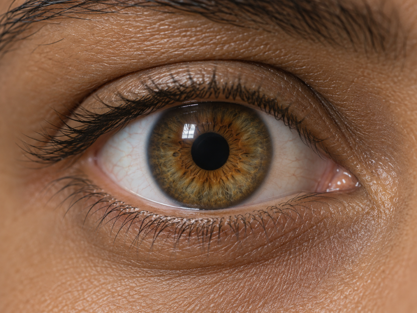
01Close-up imaging + calibrated light
Look forNew pupil asymmetry · weak light response
With sufficient eye detail and calibrated light, compare pupil size and constriction. A new size difference or reduced response can prompt an eye or neurological assessment.
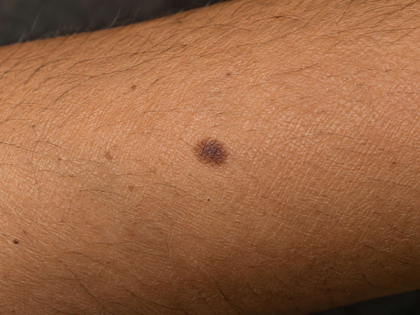
02Colour photography + depth
Look forGrowth · border or colour changes
Compare a mark’s size, borders and colour over time for dermatology review. Keep its 3D surface location; compare raised detail where the depth capture resolves it.
03RGB video analysis
Look forA resting pulse above or below your usual
Estimate pulse rate from subtle skin-colour changes in video. Persistent shifts at rest can prompt review of factors such as illness, medication or cardiovascular health.
02 / Respiration · chest movement
Use successive depth frames to track the chest as it rises and falls. Analyse breathing rate, cycle timing and differences in movement between the left and right sides.
Move over the chest → hold position → observe several breaths
01Chest depth over time
Look forUnusually fast or slow resting breathing
Count chest-rise cycles per minute. A change in resting rate can accompany illness, pain or stress; persistent departures from your usual rate deserve review.
02Left / right depth comparison
Look forUneven expansion · reduced movement
Compare each side’s chest motion. Persistent asymmetry can suggest restricted movement—from pain, chest-wall or lung problems—and prompt a respiratory assessment.
03Breathing cycle analysis
Look forPauses in chest motion · irregular cycles
Track the timing of chest movements. Repeated pauses or uneven cycles can flag a disrupted breathing pattern to discuss with a clinician.
03 / Heart + respiration · radar hold
With the arm still, radar senses tiny movements associated with breathing and heartbeat. Its module outputs include respiratory-rate and heart-rate estimates.
Pull back → settle → hold for radar acquisition
0160 GHz radar · breathing motion
Look forA sustained change in breathing rate
Read breathing-related micromotion while the arm stays still. Compare the radar’s rate estimates across sessions to highlight a persistent change that may need respiratory review.
0260 GHz radar · cardiac motion
Look forA persistently high or low resting rate
Estimate heart rate from heartbeat-related micromotion. Repeated changes from your usual resting rate give you a specific trend to discuss with your doctor.
03Multi-view colour + depth fusion
Look forChanges in visible shape and skin appearance
Combine overlapping colour and depth views across guided body positions into a textured 3D map. Revisit the same area, compare its shape and appearance over time, and share the supporting captures with your doctor.
04 / Longitudinal observation
The arm returns to park. Keep images, surface maps and signal estimates together, ready to compare with earlier sessions and review changes in your digital double.
Sequence complete · return to park
What it is
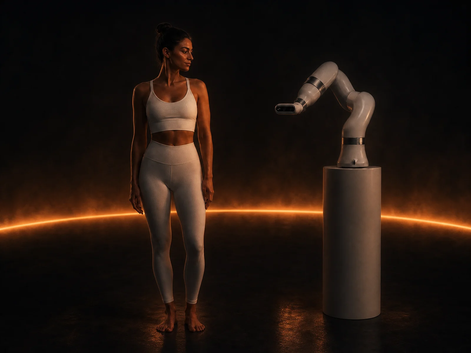
The instrument
The Bay brings the instruments to the observation. A robotic arm positions a sensor head around you, holds steady while it records, then moves to the next view. Built around repeatable routines, with room for different instruments as the system develops.
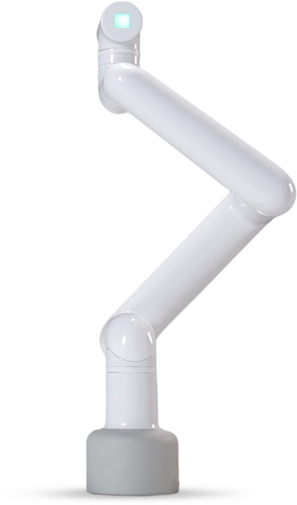
The sensing layer
3D imaging, radar and controlled light work together in one head. An integrated microphone is the next addition.
Combine colour photographs with depth to map the visible body surface. Compare contours, skin appearance and movement across scans.
Sense the tiny movements associated with heartbeat and breathing. Hold the arm still to capture heart-rate and breathing-rate estimates.
Designed to expose the same area to different wavelengths in sequence, with controlled intensity and exposure time. Compare the reflected light in each band to capture complementary views of the skin.
A microphone inside the head would add voice, breath and cough recordings. Connect sound to the same session as the images and movement.
Embodied intelligence
Guided
a planned examination routine.
Non-contact
cameras and radar at a distance.
Repeatable
return to the same observation.
Modular
one arm, different instruments.
What it does
The goal is a guided examination: the arm positions, holds for a reading and moves to the next observation. You follow the prompts; the robot follows the routine.
Cameras record visible surfaces and depth. Radar picks up the small movements associated with breathing and heartbeat while the arm holds still.
Stored viewpoints and body landmarks help line up the next observation with an earlier one, so you can compare the same area over time.
A common mounting approach makes room for different sensor heads. Start with imaging and sensing, then expand the instruments around the examination.
The arm changes its viewpoint; you change position when prompted. Front, sides and accessible back surfaces become a sequence of connected observations.
Specialist models process the images and signals. Soma connects the findings to your previous observations, and your double keeps the history.
Where it sits
The arm gathers an image, a depth map or a signal. Specialist models extract the observations; Soma reads them alongside your history and other results. Your digital double connects each finding to its source, its location and its date. One system, from examination to understanding.
The system
The integrated head
One enclosed suite moves with the wrist. The camera sees the surface, the radar follows tiny movements and the light strips shape the capture conditions. A routed cable follows the arm back to the table. Each observation keeps its viewpoint and timing, ready for Soma to connect it to the others.
A scan, in motion
Each examination follows a recorded routine: find the body landmarks, return to the viewing position, hold for the capture and check its quality. Keep the images and signals with their capture conditions, so the next session has something meaningful to compare against.
01 / position
body landmarkslocate the area
viewpoint + distancehold for capture
02 / observe
images + signalsquality check
specialist modelsextract observations
03 / connect
Somacompare with history
your digital doublerecord + explore
the source stays connected to the finding
Bring it home
Designed to bring routine observation closer to you, in the clinic and at home. A consistent examination, a connected history and observations you can take into a conversation with your doctor.
Request access →Yours · web + app
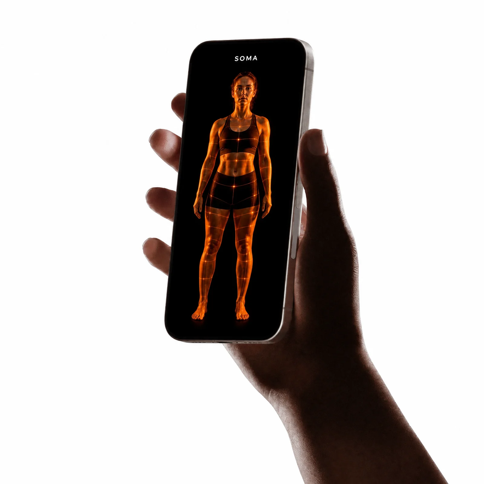
A scan from the robot. A result from the lab. A reading from your wearable. Each adds to your digital double, connected to when and where it was recorded. Soma brings the observations together, so you can follow changes over time, ask what they mean and choose what to share with your doctor.
Web + app
Your double, wherever you are.
Connect or upload
Robot scans, lab results, wearables and records you add yourself.
Always updating
Every new reading sharpens the double.
Yours, private
Encrypted, exportable — flags to discuss, never a diagnosis.
Human reference: Human Reference Atlas, HuBMAP · CC BY 4.0 · posed and simplified. Arm image: Elephant Robotics.